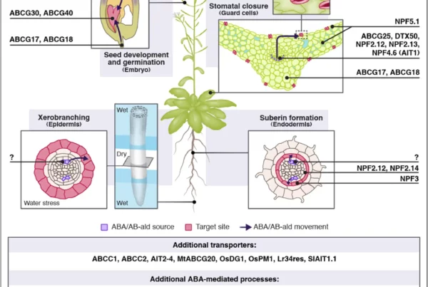
Abscisic acid activity across tissues: Linking biosynthesis, transport, and target-site action
July 23, 2026
Abscisic acid activity across tissues: Linking biosynthesis, transport, and target-site action
Anfang, M., & Shani, E. (2026). Abscisic acid activity across tissues: Linking biosynthesis, transport, and target-site action. Current Opinion in Plant Biology, 93, 102932. https://doi.org/10.1016/j.pbi.2026.102932
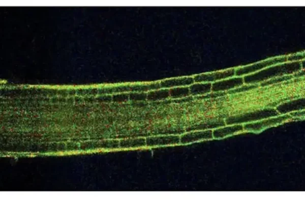
![Image: Description and validation of pMDS plasmid system for dual analysis of transcription and translation in plants. (A) Organization of pMDS1 vector showing reporters for transcription [mTurquoise (mTurQ)], translation (C-terminal mVenus), and a 2A self-cleaving peptide. (B) Organization of pMDS2 vector showing reporters for transcription (mTurQ), translation (N-terminal mVenus), and a 2A self-cleaving peptide. (C) Confocal image of pMDS1_SHRpro:SHR:mVenus:mTurQ showing gene expression (mTurQ) in the stele region and protein (mVenus) translocating to endodermis in the root meristem. (D) Confocal image of pMDS2_VAM3pro:mVenus:VAM3:mTurQ showing subcellular expression (mTurQ) in the nucleus and protein (mVenus) moving to the vacuole in the root epidermis. Red channel shows mCherry expression. nu, nucleus; vac, vacuole; *, endodermis of root meristem. Scale bar, 10 μM. Credit: Science Advances](https://hydrosensing.eu/wp-content/uploads/2026/01/sciadv.adw9153-f1-600x403.webp)